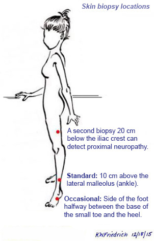
Neurodiagnostic skin biopsy has become the gold standard diagnostic test for obtaining objective pathological evidence of the presence of small-fiber neuropathy. Biopsies can be removed in almost any medical setting, often by dermatologists and some neuromuscular specialists. They are then sent to special pathology laboratories that make slides immunolabeled with a special marker (PGP9.5) that brings out the nerve endings and permits them to be counted and compared to expected numbers. Very low densities are accepted as pathological confirmation of neuropathy in clinically suspected patients. Early, mild, recovering, or patchy neuropathies may not severely damage the nerve endings in the specific skin location biopsied, so some patients with neuropathy still have a normal biopsy.
Various leading university hospital neurology departments that study peripheral neuropathy and several commercial laboratories receive and analyze these skin biopsies. Different labs use different methods to judge whether or not a skin biopsy appears normal or reveals neuropathy, so patients should ask where their skin biopsies will be sent and obtain the official copy of the test result.
For listings of doctors who perform skin biopsies for diagnosing peripheral neuropathy, see our directory of physicians in the United States or around the world.
Nerve Biopsy
This is an older technique performed only at special centers. A surgeon removes a small piece of a sensory nerve under anesthesia, usually from the lower leg, and sends it for pathological study. For many but not all patients, skin biopsy can replace nerve biopsy. Nerve biopsies remain useful for detecting inflammatory causes of neuropathies such vasculitis and sarcoidosis, and for diagnosing nerve tumors or infections such as leprosy.
What to expect after a skin biopsy
- The small wound may ooze on the day of the biopsy. To minimize oozing, keep the tight bandage on for a few hours, avoid vigorous exercise for several hours, and keep your leg up when you are sitting.
- Keep the area covered with a Band-Aid for several weeks until the scab drops off. Change the Band-Aid daily after bathing. Keep the area away from dirt; swimming is okay.
- You may experience redness and swelling for a few days at the site. Once the scab falls off after a few weeks, you may see a pink scar that will fade over the following months.
- Call your doctor if redness or swelling becomes worse or doesn’t go away, or if you see pus coming from the biopsy area.
Reference:
EFNS guidelines on the use of skin biopsy in the diagnosis of peripheral neuropathy.
Lauria, G, Cornblath, DR, Johansson, O, et al.
Eur J Neurol. 2005;12:747-758. Abstract
